
Understanding Tuberculosis and Its Impact
Tuberculosis, often called TB, is a serious infection caused by bacteria. It mainly affects the lungs, but it can spread to other parts of the body. According to the World Health Organization (WHO), millions of people worldwide get TB each year. In many countries, TB remains a major health concern. Early detection and treatment are important to stop its spread.
The Role of Chest X-rays in TB Diagnosis
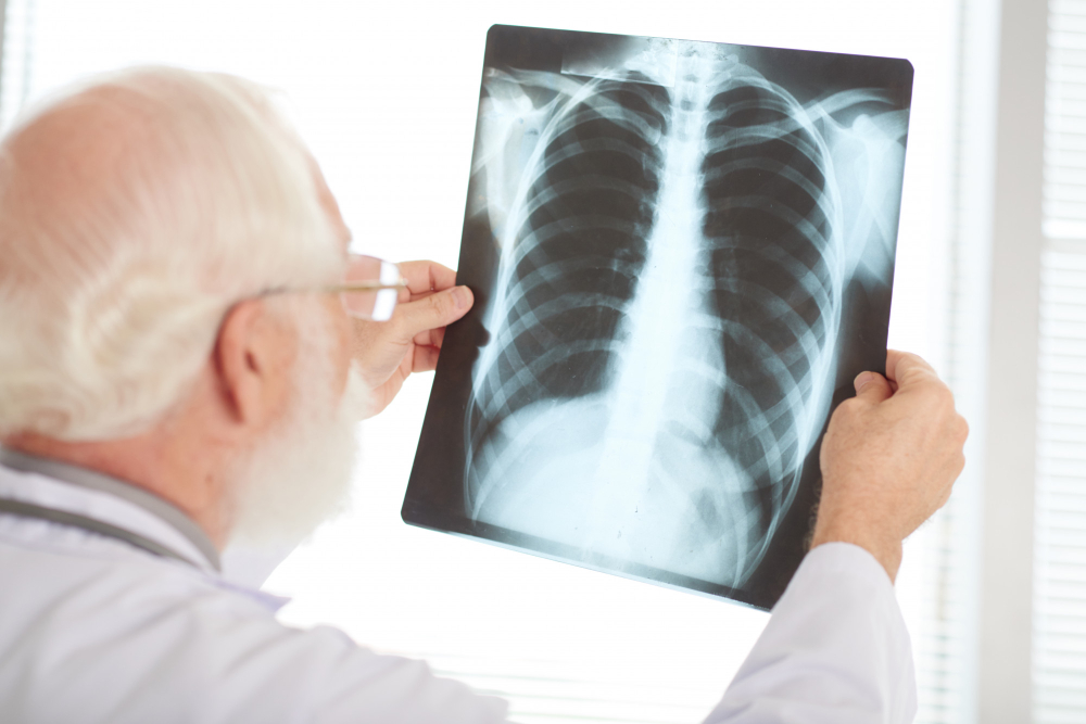
Doctors often use chest X-rays to help diagnose tuberculosis. A chest X-ray is a simple test that takes pictures of your lungs. Because TB usually starts in the lungs, this test can show changes caused by the infection. While a chest X-ray cannot confirm TB alone, it gives doctors important clues. In many places, chest X-rays are a first step in TB diagnosis imaging.
Common Chest X-ray Findings in Tuberculosis
Chest X-ray findings in tuberculosis can vary. However, some signs are more common than others. Here are the main findings doctors look for:
Additionally, TB may cause scarring or small spots called nodules. These changes can help doctors suspect TB, especially if you have symptoms like cough, fever, or weight loss.
How Radiologists Interpret Chest X-rays for TB
Radiologists are doctors trained to read X-rays. They look for patterns that match signs of tuberculosis on X-ray. For example, they check the upper parts of the lungs, where TB often starts. They also compare both lungs for differences. If they see cavities or swollen lymph nodes, they may suggest TB as a possible cause. However, radiologists use your medical history and symptoms to help make sense of the images. Sometimes, they recommend more tests for a clear answer.
Limitations of Chest X-rays in TB Diagnosis
Although chest X-rays are helpful, they have some limits. First, not all TB cases show clear changes on X-ray. Early TB or mild cases may look normal. Also, other lung problems can look like TB on X-ray. For example, pneumonia or lung cancer may cause similar shadows. Because of this, doctors often use other tests, such as sputum tests or CT scans, to confirm TB. Therefore, a chest X-ray is just one part of TB diagnosis imaging.
When to Seek Medical Attention
If you have a cough that lasts more than two weeks, you should see a doctor. Other warning signs include:
Even if your chest X-ray is normal, you should talk to a healthcare provider if you have these symptoms. Early treatment can prevent serious problems and protect others from infection.
Prevention and Early Detection Tips
Preventing TB is possible with a few simple steps. Here are some tips:
In many countries, TB screening is available for people at risk. Early detection helps stop the spread and makes treatment easier.
Summary
In summary, chest X-ray findings in tuberculosis can help doctors suspect TB. However, X-rays are only one tool in TB diagnosis imaging. If you have symptoms or abnormal X-ray results, do not wait. Consult your doctor for personalized advice and further testing. Early action can save lives and protect your community.